Forest, Herman Silva. 1954. Handbook of Algae. Knoxville, TN: University of Tennessee Press. 467 pg.
Microscopy-UK. [Internet]. Microscopy-UK, Micscape Magazine; c 1995-2011 [cited November 15, 2011]. Available from: http://www.microscopy-uk.org.uk/index.html?http://www.microscopy-uk.org.uk/pond/index.html
Microscopy-UK. [Internet]. Microscopy-UK, Micscape Magazine; c 1995-2011 [cited November 15, 2011]. Available from: http://www.microscopy-uk.org.uk/mag/indexmag.html/http://www.microscopy-uk.org.uk/mag/wimsmall/small.html
Patterson, D.J., Hedly S. 2003. Free-Living Freshwater Protozoa: A Colour Guide. Washington D.C.: Manson Publishing Ltd. 223 p.
Pennak, Robert W. 1989. Fresh-water Invertebrates of the United States: Protozoa to Mollusca. New York: Wiley.
Tuesday, November 15, 2011
Final Observation, November 10, 2011
At this final observation, the water level is very low. The aquarium is drying out. Paramecium are still abundant, but not as many as last week. There are still a great number of seed shrimp, Vorticella, and diatoms. Phacus triqueter numbers have increased. Generally, there is less activity in the upper half of the tank. A difference I notice is that there is much more debris at the bottom of the tank, but most impressive is the activity in the mud. Seed shrimp are digging around, funneling material through their bodies.
It has been interesting to see the changes in my MicroAquarium over these five weeks. There have been changes in type and number of organisms, growth, and activity. This has been a chance to participate actively in true science study, experimentation, and observation.
It has been interesting to see the changes in my MicroAquarium over these five weeks. There have been changes in type and number of organisms, growth, and activity. This has been a chance to participate actively in true science study, experimentation, and observation.
Fourth Observation, November 3, 2011
When I pick up my MicroAquarium today, I can see quite a bit of waste around the plants and at the bottom mud layer. Plants A and B, identified during set-up, are not thriving. Diatoms litter the tank, literally everywhere. Paramecium species have multiplied and congregate mostly in the lower half of the tank. Colochaete irregularis is branching more, but setae are still not present. An Amoeba species seems to move in an oozing fashion (Microscopy-UK 1995-2011). One end looks like a tied balloon. A newly identified organism, Phacus triqueter is present (Forest 296). These algae look like small ovoid-shaped green leaves with a tiny stem. Each has a red eye spot. Seemingly randomly, these algae are stationary mostly in singles or pairs.


Third Observation, October 27, 2011
After the initial set-up, One Beta Food Pellet was added to the aquarium on Friday, October 21, 2011. Pellet details: "Atison's Betta Food" made by Ocean Nutrition, Aqua Pet Americas, 3528 West 500 South, Salt Lake City, UT, 84104. The ingredients are: fish meal, wheat flour, soy meal, krill meal, minerals, vitamins, and preservatives. The analysis is: crude protein 36%; crude fat 4.5; crude fiber 3.5%; moisture 8%; and ash 15%.
At this observation, I see most, if not all of the organisms previously identified, but also some new organisms. The number of organisms has increased dramatically, in part because of the addition of the food pellet.
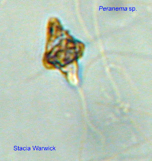
Near the soil, I count at least four of the same Paramecium species, larger than most of the other organisms (Microscopy-UK 1995-2011). These organisms are easy to identify because of the fold present. Today, the most notable organism is a flagellate with an anterior flagella pulling it along and an interesting top shaped area. With Dr. McFarland's help and much searching through the textbooks in the lab, we decide that the organism is a Peranema (Patterson 51) with either attached debris or an attached amoeba. Previously, I identified the organism as Notosolenus-like, but the flagella movement helped more closely identify it.

At this observation, I see most, if not all of the organisms previously identified, but also some new organisms. The number of organisms has increased dramatically, in part because of the addition of the food pellet.
Near the soil, I count at least four of the same Paramecium species, larger than most of the other organisms (Microscopy-UK 1995-2011). These organisms are easy to identify because of the fold present. Today, the most notable organism is a flagellate with an anterior flagella pulling it along and an interesting top shaped area. With Dr. McFarland's help and much searching through the textbooks in the lab, we decide that the organism is a Peranema (Patterson 51) with either attached debris or an attached amoeba. Previously, I identified the organism as Notosolenus-like, but the flagella movement helped more closely identify it.


Second Observation, October 20, 2011
Upon entering the lab and looking at my MicroAquarium with the unaided eye, the water level seems lower and a few air bubbles are present. Plant A seems to be darker in color. On top of the mud layer, there is a small amount of debris.
Under the microscope, there are many organisms to observe and identify. A branched algae, Coleochaete irregularis, is growing (Forest 89). Ciliates and flagellates are busy moving around the tank. One flagellate, a Notosolenus-like organism, has a long flagellum pulling it along (Patterson 54). Turning slowly, an oval-shaped ciliate passes by. I am unable to determine exactly which ciliate it is, but vacuoles and short surrounding cilia are easy to discern. Tiny clusters of diatoms are attached primarily to the sides of the tank. Many Ostracoda class seed shrimp zoom around, scavenging, and I recall that some were in the tank at the initial observation at set-up (Microscopy-UK 1995-2011). I notice several plant bladders. Inside a dead bladder there is a dead seed shrimp. Interestingly, a rotifer is feeding inside the bladder. At the mouth of another plant bladder, a Philodina species rotifer is attached to the inside by its feet (Patterson 27). Also, many Vorticella species are attached to the edges of plant bladders (Patterson 113).


Under the microscope, there are many organisms to observe and identify. A branched algae, Coleochaete irregularis, is growing (Forest 89). Ciliates and flagellates are busy moving around the tank. One flagellate, a Notosolenus-like organism, has a long flagellum pulling it along (Patterson 54). Turning slowly, an oval-shaped ciliate passes by. I am unable to determine exactly which ciliate it is, but vacuoles and short surrounding cilia are easy to discern. Tiny clusters of diatoms are attached primarily to the sides of the tank. Many Ostracoda class seed shrimp zoom around, scavenging, and I recall that some were in the tank at the initial observation at set-up (Microscopy-UK 1995-2011). I notice several plant bladders. Inside a dead bladder there is a dead seed shrimp. Interestingly, a rotifer is feeding inside the bladder. At the mouth of another plant bladder, a Philodina species rotifer is attached to the inside by its feet (Patterson 27). Also, many Vorticella species are attached to the edges of plant bladders (Patterson 113).


Monday, October 24, 2011
Initial Set-up
For my MicroAquarium, I chose water source 13, water from a plastic bird bath pool. Numerous animals have visited this site, so I thought it would be interesting to see what types of organisms were present. After adding the source water, I added two different plants: Amblestegium sp., a moss, from Carters Mill Park and Utricularia gibba L., a flowering carnivorous plant, originally from Spain Lake's south shore but grown in a water tank outside of Hesler Biology Building.
Upon first observation, I saw a few different organisms, each appearing to be single-celled. They were shaped like tiny dots, moved slowly, and appeared to be widespread in various areas of the tank. Another unidentified organism appeared to be brown, much bigger than the tiny dots, and undulated back-and-forth very quickly. This organism was very hard to follow! Also, Dr. McFarland peered into my microscope and noticed green algae near the bottom of the tank. The algae were flat-plated with tiny tails, some individual, and some in clusters.
Weekly observations will lead to identification of the microorganisms in my tank and an understanding of the changes and life processes of these tiny forms of life.
Subscribe to:
Posts (Atom)